Logo

a subcellular Atlas of Planktonic Symbioses

earth_subcellular scale

OBJECTIVES of the AtlaSymbio project

The AtlaSymbio project aims at characterizing different cell-cell interactions in the marine and freshwater plankton by visualizing their ultrastructural organization in three dimensions (e.g. morphological integration of host-symbiont, cellular architecture of symbionts, host-symbiont interface). A biobank of samples, as well as an interactive open-source atlas will be built and available for the research community, fostering new synergies and collaborations in the symbiosis field.
AtlaSymbio was funded by the Gordon and Betty Moore Foundation through the Symbiosis in Aquatic Systems Initiative. Our project involves an interdisciplinary consortium of experts in microalgae, symbiosis and electron microscopy, from Grenoble (Cell and Plant Physiology Lab-Photosymbiosis team), EMBL Heidelberg (Schwab’s group), and Roscoff (Ian Probert, marine Station, Roscoff culture collection).

Context

In aquatic ecosystems, an extraordinary diversity of single-celled organisms fuel marine food webs and regulate the atmosphere. These microorganisms have long been considered in isolation from each other in the environment, but recent awareness of the ubiquity of intimate cellular interactions, ranging from mutualistic symbioses to parasitic relationships, has led to a paradigm shift in our understanding of aquatic ecosystems. For instance, single-celled organisms can access energy (metabolites, nutrients) for their growth through these complex interactions. Environmental genomics has unveiled a wealth of genes involved in such interactions worldwide and predicted numerous associations through co-occurrence network analyses. However, there is a need to visualize these enigmatic interactions at the subcellular level to fully understand the mechanisms that take place inside cells and their impact on the ecosystem.
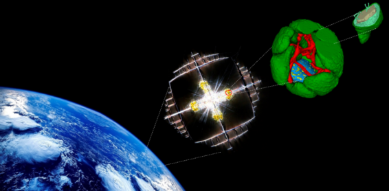
Morphological transformation of the marine microalga Phaeocystis in symbiosis unveiled by FIB-SEM: multiplication of plastids (green), extension of a mitochondrial network (red), volume increase of the nucleus (blue), including chromatin (yellow). (from Uwizeye et al PNAS 2021).
Recent progress in sample preparation and subcellular imaging techniques now offers the possibility to observe the three-dimensional structural organization of a cell within a host cell.  Focused Ion Beam Scanning Electron Microscopy (FIB-SEM) applied to samples prepared by high-pressure cryofixation can be used to shed light on structural features of symbionts (e.g. surface area and volume of energy-producing organelles) and how symbionts are structurally integrated into a host cell. Stack of images will be processed (alignment, filtering) and then semi-automatic and/or AI-based segmentation will allow to reconstruct in 3D some organelles and cellular structures of interest. Morphological changes provoked by biotic interactions can have major consequences on the bioenergetics of symbionts and thus benefits to the host. This cutting-edge 3D technique has outstanding potential to drive a step-change in our mechanistic understanding of symbiotic relationships.

AtlaSymbio will provide unprecedented open-source sample collections and datasets for the research community, which will open exciting new avenues for the study of mechanisms of cell-cell interactions in the environment.

Proposed work

Symbiotic microalgae and recent endosymbioses in culture

Microalgal species known to be symbionts (strains isolated from holobionts) and cultured holobionts (endosymbiosis/kleptoplastidy) will be prepared for 3D electron microscopy. Many symbiotic strains are available in the Roscoff Culture Collection (www.roscoff-culture-collection.org). Additional strains will be obtained from other culture collections or labs involved in the Symbiosis in Aquatic Systems Initiative.

Uncultivable marine and freshwater planktonic symbioses

The Mobile Laboratory of the TREC expedition equipped with high-pressure freezing instruments will offer a unique opportunity to prepare samples for cutting-edge subcellular microscopy from uncultivable planktonic symbioses. Sampling will be conducted at 9 sites along the European coast between Spring 2023 and Summer 2024.

Potential uncultivable symbioses targeted for AtlaSymbio involving photosymbiosis and kleptoplastidy: Radiolarians, pelagic and benthic Foraminiferans, ciliate-diatom symbioses, dinoflagellates with cyanobacteria and/or microalgae (tertiary endosymbioses), larvae of the jellyfish Vellela vellela, freshwater ciliates (Paramecium and Stentor), symbiotic Acoels, and uncultured protist models of the Symbiosis in Aquatic Systems Initiative.

Samples prepared will be either stored in liquid nitrogen or further processed for EM (i.e. resin embedded), thus building a biobank resource of specimens ready for downstream analysis. We envision to perform resin embedding and FIB-SEM imaging for some, but such vitrified specimens can also be utilized for downstream cryo-electron tomography. We will image extensively some symbioses by FIB-SEM at high isotropic resolution, and segment them to collect quantitative morphometric information. All FIB-SEM data will then be deposited on an open-source portal, in addition to protocols for sample preparation. This portal has also the ambition to foster cell biology-oriented initiatives among the community.
Light microcopy images of targeted uncultured planktonic symbioses from marine samples. A-F: radiolarians with haptophyte (Phaeocystis) or dinoflagellate (Brandtodinium) symbiotic microalgae. G-H: Pelagic foraminiferans with dinoflagellate (Pelagodinium). I: The dinoflagellate host Noctiluca with green microalgae. J: the dinoflagellate host Amphisolenia with pelagophyte symbionts.

Last news

Information of PIs

* Johan Decelle is CNRS team leader based at LPCV – Cell and Plant Physiology – laboratory (CNRS Grenoble France), working on photosymbiosis. He has developed an expertise in subcellular imaging (nanoSIMS, FIB-SEM) and single-cell transcriptomics applied to uncultivable cell-cell interactions.

* Ian Probert is a Sorbonne University research engineer based at the Roscoff marine station (Brittany, France) where he is the director of the Marine Biological Resource Centre. He manages the Roscoff Culture Collection (www.roscoff-culture-collection.org) which is the largest and most diverse public culture collection of marine organisms in the world, offering access to ca. 6000 strains of marine microalgae, cyanobacteria, bacteria, viruses and macroalgae. The research focus of the RCC team is on taxonomic description of marine microorganisms. Ian Probert isolated and cultured many microalgal strains in the past from planktonic and benthic symbioses.

* Yannick Schwab is a team leader in the Cell Biology and Biophysics at the European Molecular Biology Laboratory (EMBL) in Heidelberg, Germany. The Schwab team is specialized in developing methods for the correlation of multiple imaging modalities with volume Electron Microscopy, with a wide range of applications in the Life Sciences. Yannick Schwab is also heading the Electron Microscopy Core Facility at EMBL, that is providing access to advanced methods in cellular EM, gathering a large spectrum of techniques from sample preparation to image analysis.

Collaborative partners:

Pierre-Henri Jouneau: IRIG CEA Grenoble)

Guy Schoehn and Benoit Gallet: Structural Biology Institute, CNRS CEA Grenoble  (MEM electron microscopy platform and cryoimaging).

logo-rcc-lifting-v2